
Research Interest
RADIATION and CANCER RISK
Studies in the Pluth lab have an overall focus on investigating the cellular response to radiation and its influence on cancer risk in human cells. To this end, we study how radiation effects are temporally regulated and their dependence on dose, dose rate, cell type and radiation quality (high vs low linear energy transfer). In addition, given that the primary lesion induced by ionizing radiation is a double strand break, in examining cellular effects induced by ionizing radiation we are also investigating double-strand break repair and the network of pathways important in damage response.
CANCER RISK IN MAMMARY CELLS
The female breast is known to be highly susceptible to the cancer-causing effects of radiation, thus one tissue type we are particularly interested in investigating the effects of radiation is mammary epithelial tissue. We address these questions using both normal primary mammary fibroblast and epithelial strains and various other lines developed by our collaborators at LBNL, Drs Martha Stampfer and James Garbe (http://www.lbl.gov/~mrgs/mindex.html), which contain characteristic changes to mimic alterations that occur during cancer progression. In these studies, we investigate cell type differences in phospho-protein signaling both at early timepoints (<24h), as well as much later timepoints (weeks) following damage in mammary epithelial and mammary fibroblast cells. In addition, by measuring surrogate cancer endpoints of telomere length changes and centrosome defects in these same cell types, we are able to define how variations in the phospho-protein signaling patterns may be linked with changes associated with an increased cancer risk.
SIGNALING IN 3D ESOPHAGEAL EPITHELIUM
Another tissue which has been shown to have an increased risk for radiation induced cancer is the esophagus. In this work we study radiation effects both in 2D monolayer cultures as well as in a 3D organotypic cultures, to determine how cells respond uniquely based on growth in a 2D or 3D platforms. The overall goal of this work is to use a systems biology approach to mathematically and experimentally describe how low doses of low and high LET radiation, influence the cross-talk between the ATM and TGFb pathways. Both the ATM and TGFbpathways are activated upon radiation exposure and there is some evidence that these pathways may influence one another, but more work is needed to clearly define these interactions. In these studies we are characterizing ATM and TGFb mediated signaling in esophageal epithelial and fibroblast cells using 2D models and comparing results to the signaling patterns observed in 3D organotypic cell culture models to elucidate possible cell type and context specific differences in inter-cellular communication.
EFFECT OF RADIATION ON STEM /PROGENITOR CELLS
Recent studies have determined that changes in the stem/progenitor niche within a tissue may be particularly important in cancer development. Evidence also suggests specific sub-populations of stem-like cells in cancers are responsible for driving and maintaining growth in some tumors. These cells have been shown to be relatively resistant to radiation and other damage inducing agents commonly used in cancer therapy. Thus, it is important to understand the mechanisms of radio resistance in these cells to better understand their potential contribution to increased cancer risk as a consequence of radiation exposure. We are interested in investigating the effects of radiation in activating dormant initiated cells isolated from both esophageal and mammary tissue and identifying markers unique to these populations showing more resistance to radiation.
- Hu, S., Pluth, J.M., Cucinotta, F.A., Putative binding modes of Ku70-SAP domain with double strand DNA:a molecular modeling study. Accepted Journal of Molecular Modeling, August 2011.
- Wang, M., Hada, M., Huff, J., Pluth, J.M., Anderson, J., O’Neill, P., Cucinotta, F.A., Heavy ions can enhance TGFb mediated epithelial to mesenchymal transition. Accepted to Journal of Radiation Research August 2011.
- Chappell, L.J., Whalen, M.K., Gurai, S., Ponomarev, A., Cucinotta, F.A., Pluth, J.M. (2010), Analysis of flow cytometry DNA damage response protein activation kinetics after exposure to xrays and high-energy iron nuclei. Radiation Research 174 (6) 691-702.
- Costes, SV., Chiolo, I., Pluth, JM., Barcellos-Hoff, MH., Jakob, B. (2010), Spatiotemporal characterization of ionizing radiation induced DNA damage foci and their relation to chromatin organization. Mutat. Res., 2010 Jan 8 [Epub ahead of print].
- George, K.A., Hada, M., Jackson, L., Elliot, T., Kawata, T., Pluth, J.M., Cucinotta, F.A. (2009), Dose Response of Gamma and Iron Nuclei for Induction of Chromosomal Aberrations in Normal and Repair-Deficient Cell Lines. Radiation. Research 171 (6) 752-63.
- Whalen, M. Gurai, S., Zahed-Kargaran H., and Pluth, J.M. (2008), Specific ATM-Mediated Phosphorylation Dependent on Radiation Quality. Radiation Research, 170:353-364.
- Cucinotta, F.A., Pluth, J.M., Anderson, J.A., Harper, J.V., and O’Neill, P. (2008). Biochemical Kinetics Model of DSB Repair and Induction of gH2AX Foci by Non-homologous End Joining. Radiation Research, 169(2):214-22.
- Pluth, J.M.,Yamazaki, V, Cooper, B.A., Rydberg, B.E., Kirchgessner, C.U. and Cooper, P.K. (2008). Nijmegen Breakage Syndrome Cells Are Defective in Fidelity of DNA Double Strand Break Rejoining. DNA Repair 7(1):108-18.
- Shimura T, Martin MM, Torres MJ, Gu C, Pluth JM, DeBernardi A, McDonald JS, Aladjem MI. (2007). DNA-PK is involved in repairing a transient surge of DNA breaks induced by deceleration of DNA replication. J Mol Biol. 367(3):665-80.
- Wang, J., Pluth, J.M., Cooper, P.K., Cowan, M.J., Chen, D.J., Yannone, S.M. (2005). Artemis deficiency confers a DNA double-strand break repair defect and Artemis phosphorylation status is altered by DNA damage and cell cycle progression. DNA Repair, 4(5):556-70.
- Pluth, J.M., Fried, L.M., Kirchgessner, C. (2000). Severe combined immunodeficient cells expressing mutant hRAD54 exhibit a marked DNA double-strand break repair and error prone chromosome repair defect. Cancer Research, 61(6):2649-55.
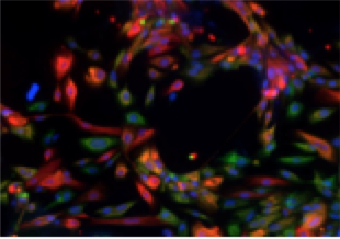
Human mammary epithelial cell line showing cells of various lineages based on keratin markers. Cells are counter-stained with DAPI (blue), keratin 8 (green), and keratin 14 (red). Cells exhibiting both K14 and K8 surface markers are visualized in yellow.
Department:
Cancer and DNA damage responses
Berkeley Lab
One Cyclotron Road
Mailstop: 977
Berkeley, CA 94720
Tel: (510)486-5799
Email: jmpluth@lbl.gov
LAB MEMBERS
Principal Scientist
Pluth, Janice
Postdoctoral Fellow
Sridharan, Deepa
Research Assistants
Whalen, Mary
Wilson, Wade
Students
Chan, Ramey
Guo, Monica
Huang, Alex
Roley, Jason
Roppel, Ryan
Administrative Assistant
Harris, Jasmine
Selected Publications
